Short answer: the evidence does not support it. In the one experiment that tested the question most directly — genetically diabetic db/db mice — keratinocyte migration did not differ from control, even though other repair endpoints improved.3 That result sits awkwardly beside a genuinely strong laboratory reputation: TB-500 and the parent molecule it derives from, thymosin beta-4, accelerate the movement of keratinocytes, endothelial cells, and fibroblasts in culture and in several non-diabetic animal wound models.12 Below we set out where that migration signal is strong, where it disappears, what the human evidence gap looks like, and what would actually settle the question. Dosing conventions used across this research are collected in our TB-500 5 mg vial protocol.
The distinction matters because “promotes cell migration in a dish” and “restores the impaired migration that defines a diabetic non-healing wound” are not the same claim, and the evidence supporting the first is far stronger than the evidence supporting the second. TB-500 is a research peptide. It is not approved by the U.S. Food and Drug Administration or any comparable regulator for treating diabetic ulcers or any other condition, its wound-healing data are overwhelmingly animal and in vitro, no human randomized controlled trial has demonstrated efficacy in diabetic wounds, and it is prohibited in sport by the World Anti-Doping Agency.11 Nothing here should be read as suggesting it treats, cures, or prevents diabetic ulcers or any disease.
This piece is written for researchers and scientifically literate readers who want an accurate map of what is known, what is merely plausible, and what is simply absent. We will work through the molecule’s identity and the naming confusion around it, the actin-based mechanism that gives it its migratory reputation, why diabetic wounds fail to migrate in the first place, what the pivotal db/db experiment actually reported, the broader in vitro and non-diabetic evidence, how it compares across models, the diabetic-adjacent findings in nerve and cornea, the disappointing human trials, research methodology, safety and sourcing, and the limitations that keep the title’s question genuinely open.
What TB-500 Actually Is — and the Naming Problem
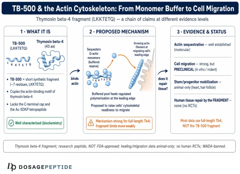
Precision about the molecule is the first casualty in most discussions of TB-500, so it is worth slowing down. Thymosin beta-4 (Tβ4) is a naturally occurring 43-amino-acid peptide, one of the most abundant intracellular proteins in mammalian cells and the principal G-actin–sequestering peptide in the cytoplasm.2 It is not a hormone in the classical sense despite the “thymosin” label, which is a historical artifact from its original isolation from thymic tissue. Its dominant cellular job is to bind monomeric (globular) actin and hold it in a reserve pool, a function that turns out to be central to how cells crawl.
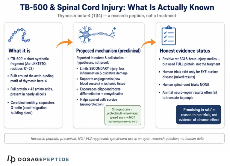
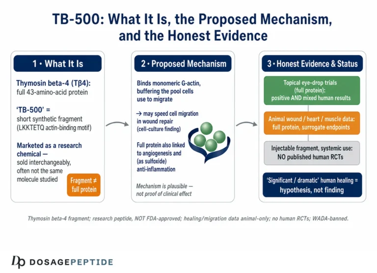
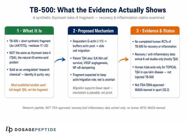
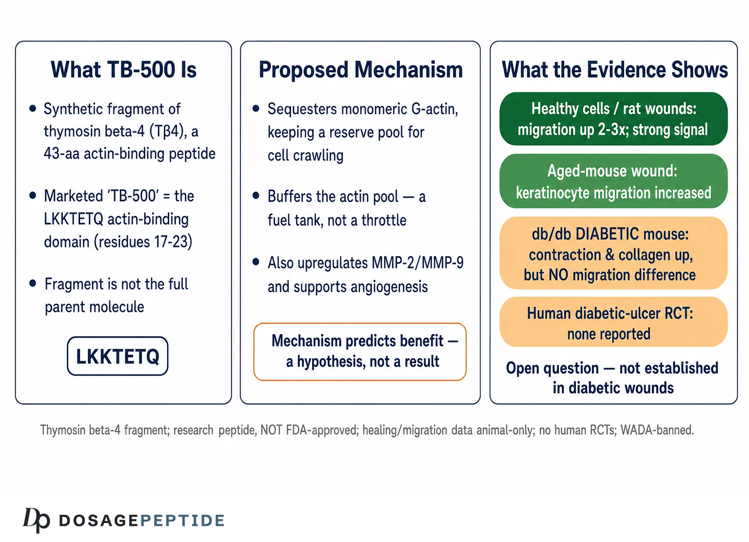
“TB-500” is where the confusion begins. In the research-chemical and athletic markets, the name is used loosely and inconsistently. Sometimes it refers to synthetic, full-length thymosin beta-4. More often, and more accurately, TB-500 denotes a shorter synthetic peptide built around the actin-binding domain of Tβ4 — the seven-residue sequence LKKTETQ, corresponding to residues 17 through 23 of the parent molecule, frequently supplied in an acetylated form.3 This fragment was chosen precisely because that heptapeptide carries much of the actin-binding and cell-migratory activity of the whole molecule, as we will see. But a fragment is not the parent. The full Tβ4 sequence has additional binding surfaces and regulatory interactions — with actin polymerization dynamics, with stem-cell and anti-inflammatory signaling — that a seven-residue peptide cannot fully reproduce.
This distinction is not pedantry; it directly undermines a great deal of the confident writing about TB-500. The overwhelming majority of the peer-reviewed wound-healing literature was generated using full-length thymosin beta-4, often the recombinant or synthetic 43-mer developed as a pharmaceutical (for example, the clinical candidate RGN-137).9 When a vendor page cites “TB-500 wound-healing studies,” it is almost always borrowing data collected on the parent peptide and attaching it to the fragment. Sometimes that borrowing is reasonable — the LKKTETQ domain does reproduce key activities — and sometimes it is not, because the fragment and the full molecule have never been compared head-to-head in the specific model being invoked. A disciplined reader keeps three objects distinct: the endogenous protein (Tβ4, 43 residues), the actin-binding domain (LKKTETQ, the source of much migratory activity), and the marketed research chemical (“TB-500,” variable in composition and purity). Collapsing these three into one is the single most common error in the popular literature, and it inflates the apparent evidence base for the fragment specifically.
For orientation within the broader research landscape, this compound sits in the same conceptual neighborhood as other so-called regenerative or repair peptides, and the site’s discussion of whether TB-500 is a promising therapy for chronic inflammatory conditions traces the same molecule through a different question. The key point to carry forward is that the migratory reputation belongs, in the primary literature, mostly to full-length thymosin beta-4, and the diabetic-wound question inherits all the caveats of that provenance.
The Cell-Migration Mechanism: Actin Sequestration and the LKKTETQ Domain
The reason thymosin beta-4 is discussed in the context of cell migration at all comes down to a single elegant piece of cell biology: the actin cytoskeleton. Directed cell migration — the crawling of a keratinocyte across a wound bed, of a fibroblast into granulation tissue, of an endothelial tip cell during angiogenesis — is fundamentally a problem of controlled actin assembly and disassembly. A migrating cell extends a leading edge (the lamellipodium) by rapidly polymerizing actin filaments, then contracts its rear. To do this quickly and repeatedly, the cell needs a large, readily mobilizable reserve of unpolymerized (monomeric, or G-) actin that it can draw on and recharge.
Thymosin beta-4 is the buffer that holds that reserve. By binding G-actin in a roughly one-to-one complex, it sequesters monomers, preventing their spontaneous polymerization while keeping them available for regulated release at the leading edge.2 In this framing, Tβ4 does not push a cell to move so much as it maintains the readiness of the machinery that makes movement possible — it keeps the actin pool primed. When extra Tβ4 is supplied to cells, the hypothesis is that this enhanced buffering capacity supports faster, more sustained cytoskeletal turnover and therefore faster migration. Critically, the actin-binding activity localizes substantially to the LKKTETQ domain, which is exactly why that heptapeptide became the basis for the TB-500 fragment.3
It helps to be concrete about what “sequestration” buys a cell. Free G-actin monomers will spontaneously nucleate and polymerize whenever their concentration climbs above a low critical threshold, which would leave a cell with filaments forming everywhere and no controllable reserve. Thymosin beta-4 holds monomers in a soluble, polymerization-incompetent complex, effectively raising the concentration of actin a cell can carry without triggering runaway assembly. When a signal arrives at the leading edge, the cell can then release monomers locally — handing them off to profilin and the nucleating machinery — to build a lamellipodium exactly where it is needed. The peptide is thus less an accelerator pedal than a fuel tank: it does not itself steer migration, but it determines how much actin substrate is available to be steered. This framing matters for the diabetic question because if the defect in a diabetic keratinocyte lies downstream of monomer availability — in the signaling that decides where and when to release actin, or in the integrin adhesions that transmit traction — then enlarging the reserve pool may do little. Supplying more fuel does not fix a broken throttle.
The migratory story does not end at actin. Thymosin beta-4 has been shown to upregulate matrix metalloproteinases during wound repair — enzymes such as MMP-2 and MMP-9 that remodel the extracellular matrix and clear a path for cells to move through.4 Migration through tissue requires not only an internal motor (the actin engine) but also the dissolution of the surrounding matrix ahead of the cell, and Tβ4 appears to touch both. It has additionally been characterized as pro-angiogenic: the same actin-binding domain that supports migration was shown to promote endothelial-cell migration and blood-vessel formation, an activity mapped directly to the LKKTETQ region in a 2003 study.2 Because a healing wound needs new vasculature to supply migrating and proliferating cells, this angiogenic arm is mechanistically intertwined with the migratory one.
On paper, then, the mechanism looks almost tailor-made for wound healing: a peptide that buffers the actin pool to enable migration, remodels matrix to open a path, and drives the angiogenesis that feeds the process. This mechanistic elegance is precisely why TB-500 attracts so much attention, and why the intuition that it “should” help diabetic wounds is so seductive. But an intuition built from healthy-cell biology is a hypothesis, not a result, and the diabetic wound is a hostile environment where several of these steps are independently broken. Whether adding an actin-sequestering peptide can overcome that hostile biochemistry is an empirical question — and, as the next sections show, the answer in the most relevant model is not the clean “yes” the mechanism might predict. Readers interested in how a related repair peptide is argued to drive the vascular side of this process can compare the discussion of what evidence supports KLOW peptides in angiogenesis and tissue repair, which examines the same angiogenesis–migration coupling from a different compound’s vantage point.
Why Diabetic Wounds Fail to Migrate in the First Place
To judge whether TB-500 could rescue migration in a diabetic non-healing wound, one has to understand why those wounds stall. A diabetic foot ulcer is not simply a normal wound that is slow; it is a wound trapped in a self-perpetuating pathological state, and cellular migration is one of the specific steps that breaks down.
Chronic hyperglycemia poisons wound repair through many convergent routes. There are over a hundred documented physiological abnormalities in diabetic wound healing, spanning impaired growth-factor signaling, defective angiogenesis, neuropathy that removes protective sensation, poor perfusion, and dysregulated inflammation that will not resolve.5 Within that thicket, the cell-migration defect is well characterized. Keratinocytes at the wound edge, which must migrate to re-epithelialize the surface, show reduced motility in a high-glucose environment, associated with altered integrin expression and disrupted signaling pathways that normally coordinate the migratory program.5 Diabetic fibroblasts are similarly crippled: in a classic comparison, fibroblasts from diabetic sources migrated less than non-diabetic controls and, tellingly, failed to increase their migration in response to hypoxia — a cue that normally mobilizes repair cells — indicating that the sensing machinery itself is impaired, not merely the effector.6
Layered on top of the migration deficit is a matrix problem. Chronic diabetic wounds tend toward a proteolytically hostile, over-inflamed environment with a disordered balance of matrix metalloproteinases and their inhibitors, so that matrix that should scaffold migrating cells is instead degraded chaotically. This is important because it complicates any naive reading of the Tβ4 mechanism: if Tβ4’s benefit in healthy wounds partly comes from raising MMP expression,4 then in a diabetic wound already suffering from dysregulated, excessive proteolysis, more MMP activity is not obviously helpful and could in principle be counterproductive. The same intervention can have opposite valence depending on the baseline state of the tissue.
The upshot is that a diabetic non-healing wound is defined, in part, by an intrinsic failure of cellular migration that arises from the metabolic state of the cells themselves. There is also a temporal dimension that reductionist assays miss entirely: a diabetic ulcer is a chronic wound stuck in a prolonged inflammatory phase, senescent and unresponsive, whereas the animal and in vitro migration studies almost always use acute, freshly created wounds that are primed to heal. Adding a pro-migratory peptide to a fresh wound that is already mobilizing its repair program is a fundamentally different intervention from coaxing migration out of a months-old, senescent, biofilm-laden ulcer bed. A migration-promoting peptide would have to overcome cell-autonomous defects — hyperglycemia-driven signaling failures inside the keratinocyte and fibroblast — not merely supply a permissive external nudge. That is a much taller order than accelerating migration in healthy cells, and it is the reason the healthy-model evidence cannot simply be transplanted to the diabetic question. The site’s examination of whether GHK-Cu could improve outcomes in chronic non-healing wounds works through the same disease context for a different peptide and is a useful companion for understanding just how resistant these wounds are to intervention.
The Pivotal Experiment: What the db/db Diabetic-Mouse Study Actually Found
The most directly relevant piece of evidence to the title’s question is a 2003 study by Philp and colleagues, working in the laboratory tradition that produced most of the foundational thymosin beta-4 wound data.3 The design is exactly the kind we want: full-thickness dermal wounds created in db/db diabetic mice (a leptin-receptor–deficient model of type 2 diabetes with genuinely delayed healing) and, in parallel, in aged mice, treated with thymosin beta-4 — and, importantly, with the LKKTETQ synthetic actin-binding fragment — in either saline or a hydrogel vehicle, versus vehicle controls. This is one of the few experiments that puts the migration-promoting peptide into an actual impaired-healing model and measures repair endpoints.
Here is the finding that popular summaries tend to blur. In the db/db diabetic mice, thymosin beta-4 treatment significantly increased wound contraction and collagen deposition relative to controls — real, measurable improvements in the repair process. But when the investigators looked specifically at keratinocyte migration, they observed no difference between treated and control diabetic animals: essentially all of the diabetic mice, treated or not, showed nearly complete wound coverage by day 8.3 In other words, in the diabetic model, the endpoint most central to the title’s question — migration — did not separate from control, while the benefit that did appear came through contraction and matrix deposition.
The contrast with the aged mice in the same study is illuminating. There, thymosin beta-4 did increase keratinocyte migration, along with contraction and collagen deposition, and the LKKTETQ fragment reproduced the parent molecule’s effect on repair.3 So the migratory effect on keratinocytes was demonstrable in one impaired model (aging) but not detectable as a treatment difference in the diabetic model. The most likely mundane explanation is that the particular db/db wound assay reached near-complete epithelial coverage quickly regardless of treatment, giving little room to detect a migration difference — a ceiling effect rather than proof of no biological activity. But that caveat cuts both ways: it means the study cannot be cited as clean evidence that TB-500 promotes migration in diabetic wounds, because in that model it did not produce a measurable migration advantage. The honest reading is that Tβ4 improved some aspects of diabetic wound repair (contraction, collagen) without a demonstrated effect on the migration endpoint itself.
This is why the framing of the title deserves scrutiny. The strongest direct experiment does not straightforwardly answer “yes.” It answers: “thymosin beta-4 improved repair in diabetic mice, but the improvement was not attributable to a measured increase in keratinocyte migration in that model, whereas it was in aged mice.” For a compound whose entire mechanistic reputation rests on migration, that is a genuinely important nuance, and one that responsible communication must preserve rather than sand away.
| Endpoint in db/db diabetic mice | Effect of thymosin beta-4 vs control |
|---|---|
| Wound contraction | Significantly increased3 |
| Collagen deposition | Significantly increased3 |
| Keratinocyte migration | No detectable difference; near-complete coverage in all animals by day 83 |
| LKKTETQ fragment (the “TB-500” sequence) | Reproduced parent-peptide repair effect in aged mice; diabetic-specific fragment data thinner3 |
The In Vitro and Non-Diabetic Migration Evidence
If the diabetic-specific migration evidence is equivocal, the general migration evidence — in healthy cells and non-diabetic wounds — is much more robust, and honesty requires giving it full weight too. This is the body of work that legitimately earns thymosin beta-4 its migratory reputation.
The foundational rat study established that adding thymosin beta-4, topically or systemically, to full-thickness dermal wounds increased re-epithelialization by roughly 42% over saline controls at day 4 and by as much as 61% at day 7, with increased collagen deposition and angiogenesis in treated wounds.1 In the cell-culture arm of that and subsequent work, thymosin beta-4 stimulated keratinocyte migration in a Boyden-chamber assay by two- to three-fold over control, with activity detectable at strikingly low quantities — on the order of picograms added to the chamber.1 That potency in a controlled migration assay is the strongest single line of evidence that the peptide acts on the migratory machinery directly, and it is consistent with the actin-sequestration mechanism.
The angiogenesis evidence reinforces the picture. Mapping studies localized the pro-angiogenic and endothelial-migratory activity to the actin-binding domain, demonstrating that the LKKTETQ region promotes endothelial-cell migration and vessel formation — a direct link between the fragment sequence marketed as TB-500 and a migratory, vascularizing effect.2 The MMP work adds the matrix-remodeling dimension, showing several-fold upregulation of MMP-2 and MMP-9 in the days after wounding.4 And engineered variants have pushed the effect further: a dimeric thymosin beta-4 construct designed to present two actin-binding domains accelerated wound healing beyond the monomeric peptide in a rodent model, offering a proof-of-concept that the migratory/angiogenic activity scales with the actin-binding motif.13
Taken together, this literature supports a defensible statement: in healthy cells and non-diabetic wound models, thymosin beta-4 (and, to the extent tested, its LKKTETQ fragment) promotes the migration of keratinocytes and endothelial cells and accelerates wound closure. What it does not license is the automatic extension of that statement to diabetic non-healing wounds, for the reasons already laid out — the disease breaks the very cellular machinery these assays assume is intact. The gap between “promotes migration in a Boyden chamber of healthy keratinocytes” and “restores migration in a hyperglycemia-damaged wound bed” is exactly the gap the title glosses over. For readers weighing the compound’s non-diabetic repair claims, the site’s review of what evidence shows TB-500 effectiveness in tendon and ligament repair post-injury examines a parallel musculoskeletal claim with the same “strong in animals, unproven in humans” shape.
Comparing the Models: Where the Migration Signal Is Strong and Where It Is Absent
Placing the evidence side by side, by model type, makes the pattern legible. The signal is strong precisely where the cells are healthy and weakens or disappears as the model approaches the clinical reality of a diabetic non-healing wound.
| Model | Cell-migration finding for Tβ4 / TB-500 | Evidence quality & relevance |
|---|---|---|
| Boyden-chamber keratinocyte assay (healthy cells) | 2–3× increase in migration at picogram doses1 | Controlled, mechanistic; low disease relevance |
| Endothelial migration / angiogenesis assays | LKKTETQ domain promotes endothelial migration and vessel formation2 | Mechanistic; supports vascular arm |
| Rat full-thickness dermal wound (healthy) | Re-epithelialization up 42–61%; increased angiogenesis1 | In vivo but non-diabetic |
| Aged-mouse wound (impaired healing) | Keratinocyte migration increased; fragment reproduced effect3 | Impaired model; migration signal present |
| db/db diabetic-mouse wound | No detectable difference in keratinocyte migration; contraction/collagen improved3 | Most relevant model; migration endpoint not met |
| Human diabetic-ulcer RCT | None specific; dermal-ulcer trials in other wound types failed to reach significance79 | Human evidence gap |
The gradient is unmistakable and, frankly, humbling for the strong version of the title’s claim. As the model moves from an isolated healthy cell in a chamber toward the actual pathology of a diabetic ulcer, the clean migration signal fades. This is a common and sobering pattern in translational biology: effects that are large and reproducible in reductionist systems attenuate or vanish as the messy, redundant, damaged reality of chronic disease is layered back in. It does not mean TB-500 does nothing — the diabetic-mouse contraction and collagen effects are real — but it means the specific proposition “promotes cellular migration in diabetic non-healing wounds” is not something the current evidence establishes.
Diabetic-Adjacent Evidence: Nerve and Cornea
Two lines of research deserve mention because they are frequently cited — sometimes over-cited — as support for TB-500 in diabetes, and it is worth being precise about what they do and do not show.
The first is diabetic peripheral neuropathy. In a study using type 2 diabetic (db/db) mice, thymosin beta-4 treatment improved measures of peripheral-nerve function, increased functional vascular density and regional blood flow in the sciatic nerve, and modulated the angiopoietin-1/angiopoietin-2 axis, with effects linked to PI3K/Akt signaling.10 This is a legitimate, peer-reviewed finding of benefit in a diabetic complication. But it concerns nerve and its microvasculature, not cutaneous wound re-epithelialization, and the primary readouts were vascular and neurological rather than keratinocyte or fibroblast migration in a wound bed. It supports a general theme — that Tβ4 has pro-vascular, tissue-protective activity in a diabetic context — without directly answering the wound-migration question. Borrowing it as evidence for diabetic ulcer healing is an extrapolation across tissue and endpoint.
The second is the eye. Thymosin beta-4 has been studied in corneal wound healing and dry-eye/ocular-surface disease, including diabetic keratopathy contexts, and reached clinical testing as an ophthalmic candidate. These studies generally support pro-migratory, anti-inflammatory, and wound-closing activity on the corneal epithelium. Again, though, the cornea is an avascular, immunologically privileged, highly specialized epithelium, and its wound biology differs substantially from a full-thickness diabetic foot ulcer with its vascular, neuropathic, and infective complexities. Positive corneal data are encouraging for the molecule’s general regenerative profile but are not a substitute for diabetic dermal-wound evidence.
The honest synthesis of the diabetic-adjacent literature is that thymosin beta-4 shows tissue-protective and pro-vascular activity in more than one diabetic complication model, which makes the wound hypothesis reasonable to pursue — but each of these findings sits in a different tissue with different endpoints, and none of them closes the specific gap around cutaneous cellular migration in diabetic non-healing wounds.
The Human Evidence Gap
For all the animal and mechanistic work, the clinical record is where the strong version of the title’s claim runs out of road. There is no published randomized controlled trial demonstrating that thymosin beta-4 — let alone the TB-500 fragment specifically — heals diabetic foot ulcers or measurably restores cellular migration in human diabetic wounds. What human dermal-wound data exist come from adjacent chronic-wound indications, and they are, at best, inconclusive.
Thymosin beta-4, formulated for topical dermal delivery as the pharmaceutical candidate RGN-137, was taken into Phase 2 trials for chronic wounds. In a European prospective, randomized, placebo-controlled dose-escalation study in venous stasis ulcers, thymosin beta-4 was reported to be safe and well tolerated, with signals suggesting enhanced healing at particular doses.78 But when the compound was carried into larger, blinded, placebo-controlled Phase 2 dose-response trials in pressure ulcers and venous stasis ulcers, the results were sobering: the drug was again safe and well tolerated, but there were no statistically significant differences between placebo and any RGN-137 dose for complete wound healing or rate of healing. The most that could be said was that a mid-dose arm appeared to initiate healing somewhat more rapidly, an observation that did not reach statistical significance.9
It is worth pausing on why a “safe but not significantly effective” result is so common in chronic-wound trials, because it bears on how to read the thymosin beta-4 data specifically. Chronic-wound trials are notoriously difficult: the wounds are heterogeneous in size, depth, duration, and cause; the standard of care against which any drug competes — debridement, off-loading, compression, moisture control, infection management — is itself effective enough to close a substantial fraction of wounds on its own, compressing the room a drug has to show benefit; and placebo groups in well-run trials often heal surprisingly well precisely because trial enrollment improves adherence to that standard of care. Against that backdrop, a true but modest pharmacological effect can easily be swamped, and a compound can be both genuinely active in a dish and undetectable in a trial. This does not rescue thymosin beta-4 — a drug that cannot demonstrate benefit above good wound care is, for practical purposes, unproven — but it does mean the negative trials should be read as “failed to demonstrate efficacy” rather than “proven inert.” The distinction is small comfort clinically and large scientifically.
Several honest conclusions follow. First, even in chronic wounds that are not diabetic — venous and pressure ulcers — the human efficacy signal for thymosin beta-4 failed to separate convincingly from placebo. Second, diabetic ulcers specifically were not the population in which even these equivocal trials were run, so the compound’s clinical record in the exact indication implied by the title is essentially empty. Third, the trials used full-length thymosin beta-4, not the LKKTETQ “TB-500” fragment sold in research-chemical channels, so even the negative human data do not directly characterize the marketed compound. The pattern — reassuring safety, unconvincing efficacy — is a familiar one across the regenerative-peptide field and should temper any confident reading of the animal migration data.
This is the crux of responsible communication about TB-500 and diabetic wounds: the mechanism is attractive, the healthy-model data are genuinely strong, and the human data in related chronic wounds did not deliver. That combination warrants continued research interest, not clinical claims. For a broader sense of how the compound’s recovery claims fare when held to a clinical standard, the site’s look at whether clinical studies show TB-500 really speeds recovery and reduces inflammation arrives at a similarly measured verdict.
Research Models and Methodology
Understanding how this question has been studied clarifies what the data can and cannot support, and what a definitive answer would require. The methodology falls into recognizable tiers, each with characteristic strengths and blind spots.
In vitro migration assays. The workhorse is the Boyden chamber (transwell) assay, in which cells migrate through a porous membrane toward a stimulus, and the scratch/wound-closure assay, in which a cleared lane in a confluent monolayer is monitored for re-closure. These are where thymosin beta-4’s two- to three-fold migration effect on keratinocytes was demonstrated.1 Their strength is mechanistic clarity and dose control; their weakness is that they typically use healthy, often immortalized, cells under standard glucose conditions. A methodologically honest test of the diabetic question would run these assays under sustained high-glucose conditions or with cells derived from diabetic donors, and would measure not just migration distance but the specific molecular lesions hyperglycemia imposes — integrin expression, p38/MAPK signaling, autophagy — to see whether Tβ4 corrects them. Such targeted experiments are sparse.
Animal wound models. The db/db mouse is the standard genetic model of impaired diabetic healing, and the aged mouse and streptozotocin-induced diabetic rodents are also used. The Philp study’s use of db/db mice with full-thickness wounds and defined endpoints (contraction, collagen, keratinocyte migration) is methodologically appropriate.3 The limitations are the ones common to rodent wound work: mice heal substantially by contraction (via the panniculus carnosus muscle) rather than the re-epithelialization that dominates human healing, which is precisely why a rodent “contraction” benefit may not translate; wound-splinting models exist to force re-epithelialization-dominant healing but were not the design here. Ceiling effects, as likely occurred with the day-8 near-complete coverage, can also mask or manufacture apparent treatment differences.
Human trials. The RGN-137 program used the correct clinical architecture — randomized, double-blind, placebo-controlled, dose-response designs with objective wound-closure endpoints.9 Their limitation for the present question is population: venous and pressure ulcers, not diabetic foot ulcers, and full-length Tβ4 rather than the fragment. A definitive answer to the title would require a properly powered randomized controlled trial in diabetic foot ulcers, with standardized off-loading and wound care as the comparator backbone, objective closure endpoints, and ideally mechanistic sub-studies (wound-edge biopsies assessing migration markers) to connect any clinical effect back to the migration hypothesis. No such trial has been reported.
The methodological bottom line is that the evidence architecture for TB-500 in diabetic wounds is inverted relative to what the title assumes: it is strongest in the least disease-relevant systems and thinnest exactly where clinical relevance is highest. Researchers documenting handling and study parameters can find general practices summarized in the site’s peptide reconstitution guide, but no amount of methodological polish substitutes for the missing diabetic-wound trials.
Safety, Sourcing, and Regulatory Status
Because TB-500 circulates largely outside regulated medicine, its safety and regulatory picture must be stated carefully — and separated cleanly from any efficacy question, since the two are routinely conflated in marketing.
In the human chronic-wound trials, thymosin beta-4 was consistently reported as safe and well tolerated across the doses studied, with no dose-limiting or drug-related serious adverse events attributed to the compound.79 That is genuinely reassuring for short-term topical exposure in the studied populations, but it comes with the usual caveats: the trials were short, the populations were not diabetic-ulcer patients with their attendant comorbidities, the material was pharmaceutical-grade full-length Tβ4 rather than a research-chemical fragment, and “no signal in a small trial” is not the same as “established long-term safety.” A theoretical concern that recurs in discussions of any pro-angiogenic, pro-migratory peptide is oncological: mechanisms that help cells migrate and recruit blood vessels are the same mechanisms tumors exploit, and thymosin beta-4 has been studied in the context of tumor metastasis. This is a hypothesis-level concern, not a demonstrated harm, but it is a reason for caution rather than complacency.
Sourcing is a distinct and serious issue. Material sold as “TB-500” is offered as a research chemical of variable and often unverified purity, composition (fragment versus full-length), and sterility. Impurities, endotoxin contamination, incorrect sequence, and mislabeled mass are real and documented risks in the gray-market peptide supply, and they have nothing to do with the molecule’s intrinsic biology and everything to do with provenance. Any observation made with such material — even an informal one — is confounded by the possibility that the vial does not contain what the label claims.
On regulation: TB-500 / thymosin beta-4 is not approved by the FDA, the European Medicines Agency, or any comparable regulator for diabetic ulcers or any other therapeutic indication; its dermal-wound development did not yield an approval. It is prohibited in sport at all times by the World Anti-Doping Agency, which explicitly lists “Thymosin-β4 and its derivatives, e.g. TB-500” under the growth-factors category of the Prohibited List.11 For any athlete subject to testing, use is an anti-doping rule violation regardless of pharmacology. A recent scoping review of thymosin beta-4 and TB-500 across tissue-healing applications reached the same overall verdict that recurs throughout this article: promising preclinical activity, a persistent shortage of rigorous human evidence, and a need for controlled trials before any clinical claim is warranted.12
Limitations and What Would Actually Settle the Question
Drawing the threads together, the limitations bearing on the title’s question are substantial and compound one another rather than sitting in isolation.
The direct evidence is equivocal, not affirmative. The one well-designed diabetic-mouse wound study found improved contraction and collagen but no measurable keratinocyte-migration difference in the diabetic animals.3 The strong migration signal lives in healthy cells and non-diabetic or aged models, not in the diabetic model itself.
The compound identity is unstable. Most primary data concern full-length thymosin beta-4; “TB-500” usually denotes the LKKTETQ fragment; the two are not interchangeably validated in diabetic wounds. Claims about “TB-500” frequently rest on data generated with a different molecule.
The disease breaks the mechanism’s assumptions. Diabetic wounds fail to migrate because of cell-autonomous, hyperglycemia-driven defects,56 and the wound is already proteolytically dysregulated — a state in which Tβ4’s MMP-upregulating activity4 is of ambiguous benefit. A migration-promoting peptide must correct broken cells, not merely nudge healthy ones.
The human data, where they exist, are negative or inconclusive. Controlled trials in related chronic wounds did not show significant efficacy,9 and no diabetic-foot-ulcer RCT has been reported at all.
Translation across tissue and species is unreliable. Rodent healing is contraction-dominated; corneal and neural benefits do not transfer automatically to dermal ulcers; picogram potency in a chamber does not predict clinical closure.
To actually settle the question would require a coherent program: high-glucose and diabetic-donor migration assays that test whether Tβ4/LKKTETQ corrects the specific molecular lesions of hyperglycemia; splinted diabetic-rodent wound models that isolate re-epithelialization (migration) from contraction, with the fragment and full-length peptide compared head-to-head; and, ultimately, a properly powered randomized controlled trial in diabetic foot ulcers with objective closure endpoints and wound-edge mechanistic sub-studies. Until that work exists, the accurate answer to “Does TB-500 promote cellular migration in diabetic non-healing wound models?” is: the mechanism predicts it should, healthy-model data support the general capacity, but the one directly relevant diabetic model did not demonstrate a migration effect, and human confirmation is absent. That is an open question, not a settled fact. Readers tracking how the broader repair-peptide field handles this exact translational gap may find the cross-compound comparison in the discussion of whether AOD-9604 could enhance healing in diabetic foot ulcers and wound repair instructive, and the foundational overview of what BPC-157, the healing peptide, is provides useful context on how “healing peptide” claims are built and where they hold up.
Frequently Asked Questions
Does TB-500 promote cell migration in diabetic wounds?
Not conclusively. Thymosin beta-4, the parent peptide, robustly promotes keratinocyte and endothelial migration in healthy cells and non-diabetic wound models — two- to three-fold in Boyden-chamber assays and up to 61% faster re-epithelialization in healthy rat wounds.1 But in the most directly relevant experiment, db/db diabetic mice, thymosin beta-4 improved wound contraction and collagen deposition without a measurable difference in keratinocyte migration.3 So the migration effect that defines the molecule’s reputation was not demonstrated in the diabetic model itself, and no human diabetic-ulcer trial has confirmed it. The mechanism predicts benefit; the diabetic-specific evidence does not yet establish it.
Is TB-500 the same thing as thymosin beta-4?
Not exactly, and the difference matters. Thymosin beta-4 is the natural 43-amino-acid peptide. “TB-500” usually refers to a synthetic fragment built around its actin-binding domain, the seven-residue LKKTETQ sequence (often acetylated), though the name is used loosely and sometimes denotes the full peptide.3 Most published wound-healing data were generated with full-length thymosin beta-4, so claims attached to “TB-500” frequently borrow evidence from the parent molecule rather than the fragment being sold.
How is TB-500 supposed to work on migrating cells?
Thymosin beta-4 is the main cellular buffer for monomeric (G-) actin. By sequestering actin monomers, it keeps a mobilizable reserve ready for the rapid cytoskeletal assembly and disassembly that cell crawling requires, and the actin-binding activity localizes to the LKKTETQ domain.2 It also upregulates matrix-remodeling enzymes (MMP-2 and MMP-9) that clear a path for migrating cells4 and promotes the angiogenesis that supplies healing tissue.2
Why might diabetic wounds not respond even if the mechanism is sound?
Because diabetic wounds fail to heal partly through cell-autonomous defects: high glucose impairs keratinocyte migration via altered integrin expression and disrupted p38/MAPK and autophagy signaling, and diabetic fibroblasts migrate poorly and fail to respond to hypoxic cues.56 A migration-promoting peptide would have to correct these broken internal programs, not merely provide an external nudge to otherwise healthy cells — a far harder task.
Are there human trials of thymosin beta-4 in chronic wounds?
Yes, but not in diabetic ulcers, and the results were unconvincing. Topical thymosin beta-4 (RGN-137) was tested in Phase 2 trials for venous stasis and pressure ulcers. It was safe and well tolerated, but showed no statistically significant improvement over placebo in complete healing or healing rate, with only a non-significant hint of faster healing initiation at a mid dose.79 No diabetic-foot-ulcer randomized controlled trial has been published.
Does the evidence in diabetic neuropathy or corneal healing count?
It is supportive context, not a direct answer. Thymosin beta-4 improved nerve function and microvascular measures in diabetic (db/db) mice10 and has shown pro-migratory, wound-closing activity on the corneal epithelium. But these are different tissues with different endpoints from a diabetic dermal ulcer, so they cannot substitute for cutaneous diabetic-wound migration data.
Is TB-500 approved or legal to use?
It is not approved by the FDA, EMA, or comparable regulators for diabetic wounds or any therapeutic use, and its dermal-wound development did not produce an approval. It is prohibited at all times in sport by the World Anti-Doping Agency, which lists thymosin-β4 and its derivatives, including TB-500, under growth factors.11 Material sold as “TB-500” is a research chemical of variable purity and composition.
What would it take to answer the title’s question definitively?
A coherent research program: migration assays under high-glucose or diabetic-donor conditions testing whether the peptide corrects the specific hyperglycemic lesions; splinted diabetic-rodent wound models that isolate re-epithelialization from contraction, comparing the fragment and full-length peptide directly; and a properly powered randomized controlled trial in diabetic foot ulcers with objective closure endpoints and wound-edge mechanistic sub-studies.12 None of these has been reported, so the question remains genuinely open.
References
- Malinda KM, Sidhu GS, Mani H, et al. Thymosin beta4 accelerates wound healing. J Invest Dermatol. 1999;113(3):364-368. PMID: 10469335. https://pubmed.ncbi.nlm.nih.gov/10469335/
- Philp D, Huff T, Gho YS, Hannappel E, Kleinman HK. The actin binding site on thymosin beta4 promotes angiogenesis. FASEB J. 2003;17(14):2103-2105. PMID: 14500546. https://pubmed.ncbi.nlm.nih.gov/14500546/
- Philp D, Badamchian M, Scheremeta B, Nguyen M, Goldstein AL, Kleinman HK. Thymosin beta4 and a synthetic peptide containing its actin-binding domain promote dermal wound repair in db/db diabetic mice and in aged mice. Wound Repair Regen. 2003;11(1):19-24. PMID: 12581423. https://pubmed.ncbi.nlm.nih.gov/12581423/
- Philp D, Scheremeta B, Sibliss K, et al. Thymosin beta4 promotes matrix metalloproteinase expression during wound repair. J Cell Physiol. 2006;208(1):195-200. PMID: 16607611. https://pubmed.ncbi.nlm.nih.gov/16607611/
- Brem H, Tomic-Canic M. Cellular and molecular basis of wound healing in diabetes. J Clin Invest. 2007;117(5):1219-1222. PMID: 17476353; PMCID: PMC1857239. https://pmc.ncbi.nlm.nih.gov/articles/PMC1857239/
- Lerman OZ, Galiano RD, Armour M, Levine JP, Gurtner GC. Cellular dysfunction in the diabetic fibroblast: impairment in migration, vascular endothelial growth factor production, and response to hypoxia. Am J Pathol. 2003;162(1):303-312. PMID: 12507913; PMCID: PMC1851127. https://pmc.ncbi.nlm.nih.gov/articles/PMC1851127/
- Guarnera G, DeRosa A, Camerini R. Thymosin beta-4 and venous ulcers: clinical remarks on a European prospective, randomized study on safety, tolerability, and enhancement on healing. Ann N Y Acad Sci. 2007;1112:407-412. PMID: 17495250. https://pubmed.ncbi.nlm.nih.gov/17495250/
- Guarnera G, DeRosa A, Camerini R. The effect of thymosin treatment of venous ulcers. Ann N Y Acad Sci. 2010;1194:207-212. PMID: 20536470. https://pubmed.ncbi.nlm.nih.gov/20536470/
- RegeneRx Biopharmaceuticals. Study of Thymosin Beta 4 (RGN-137) in Patients With Venous Stasis Ulcers (Phase 2, randomized, double-blind, placebo-controlled dose-response). ClinicalTrials.gov Identifier NCT00832091. https://clinicaltrials.gov/study/NCT00832091
- Wang L, Chopp M, Szalad A, et al. Thymosin beta4 promotes the recovery of peripheral neuropathy in type II diabetic mice. Neurobiol Dis. 2012;48(3):546-555. PMID: 22922221; PMCID: PMC3533234. https://pmc.ncbi.nlm.nih.gov/articles/PMC3533234/
- World Anti-Doping Agency. The 2026 Prohibited List (S2.3 Growth Factors: Thymosin-β4 and its derivatives, e.g. TB-500). https://www.wada-ama.org/en/prohibited-list
- Thymosin Beta-4 and TB-500 in Tissue Healing, Regeneration, and Musculoskeletal Repair: A Scoping Review. Appl Sci. 2026;16(12):6202. https://www.mdpi.com/2076-3417/16/12/6202
- Xu TJ, Wang Q, Ma XW, et al. A novel dimeric thymosin beta 4 with enhanced activities accelerates the rate of wound healing. Drug Des Devel Ther. 2013;7:1075-1088. PMCID: PMC3792846. https://pmc.ncbi.nlm.nih.gov/articles/PMC3792846/
Educational and research-use disclaimer: This article is provided solely for scientific and educational purposes. TB-500 (a synthetic fragment of thymosin beta-4) is not approved by the FDA, EMA, or any comparable regulator for the treatment, cure, or prevention of diabetic foot ulcers, non-healing wounds, or any other disease, and no human randomized controlled trial has demonstrated efficacy for diabetic wound healing. The cell-migration, angiogenic, and wound-repair data discussed here are predominantly in vitro and animal findings, and the single most relevant diabetic-mouse model did not demonstrate a keratinocyte-migration effect. Nothing here is medical advice or a recommendation for human use. TB-500 is prohibited in sport at all times by the World Anti-Doping Agency. Any legitimate investigation of this compound should occur within properly authorized preclinical or clinical research under appropriate oversight. Readers should consult qualified professionals and applicable regulations before making any decisions.